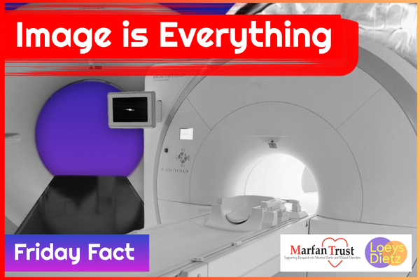
Bringing the unseen into focus, medical imaging is essential in healthcare. Today is World Radiography Day and we are celebrating the vital pieces of equipment used to facilitate Marfan and Loeys-Dietz diagnosis and care.
World Radiography Day (WRD) is celebrated on 8 November each year. The date marks the anniversary of the discovery of x-radiation by Wilhelm Roentgen in 1895.
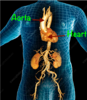
Of the many symptoms associated with Marfan and Loeys-Dietz syndromes, the most serious is a weakened aortic wall which can lead to an aneurysm and subsequent dissection. This situation is monitored carefully by medical imaging …
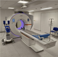
A CT (computed tomography) Scan .....
… is a test that takes detailed pictures of the inside of your body. It is an effective method and remains the most widely employed technique for defining the maximum diameter of a thoracic aortic aneurysm and monitoring the diameter over time.
Uses X-rays (radiation) to obtain 3D images of your body structures
· Fast (usually under 15 minutes)
· Contrast may be used to improve pictures
· Better for acute/emergency situations like aortic dissection
· Widely available nationwide
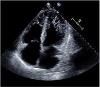
An Echocardiogram
…. or echo is a type of ultrasound scan that looks at the heart and surrounding blood vessels to diagnose and monitor various heart conditions.
A radiographer will use a probe to scan your chest, with images produced by the sound waves emitted viewed on a monitor and analysed to produce a diagnosis
· The echo looks at the heart structure e.g. pump function, valve function, and the size of the aorta
An MRI (magnetic resonance imaging)
… is a type of scan that uses strong magnetic fields and radio waves to produce detailed 3D images of the inside of the body. It has great potential in the study of the thoracic aorta and has the advantage over CT in that it is able to accurately visualise the entire aorta without using ionizing radiation and is able to give additional information on ventricular, valvular and vascular function and flow dynamics.
· It is slower than a CT scan and takes usually between 30 and 60 minutes
· Contrast may be used to improve pictures
· Better for long-term aortic monitoring and repeated imaging (due to no use of radiation)
· May need to travel to a specialist centre for detailed cardiac/aortic imaging
· Can be claustrophobic
· Need to make staff aware of any metal in the body e.g. pacemakers
An MRI is also used to diagnose dural ectasia as it is better at imaging the dural sac (Pollock et al, 2021) than other imaging techniques like computed tomography (CT) although these can be used if MRI is contraindicated.
Dural ectasia is the enlargement of the Dura. It can cause radicular pain in the buttocks and legs; nerve root paralysis affecting bowel and bladder function and motor function in the legs; and dull, unremitting discomfort in the lower abdomen and pelvis. It occurs in 63–92% of people with Marfan syndrome. Dural ectasia may also occur in Ehlers-Danlos Syndrome, neurofibromatosis type I, ankylosing spondylitis, and trauma.
